

A-P.40: Uncovering the Dark Side of the Immunopeptidome
Authors
Marcel Wacker
Department of Peptide-based Immunotherapy, Institute of Immunology, University and University Hospital Tübingen
Jens Bauer
Department of Peptide-based Immunotherapy, Institute of Immunology, University and University Hospital Tübingen
Jonas Scheid
Department of Peptide-based Immunotherapy, Institute of Immunology, University and University Hospital Tübingen
Annika Nelde
Department of Peptide-based Immunotherapy, Institute of Immunology, University and University Hospital Tübingen
Sven Nahnsen
Juliane Walz
Department of Peptide-based Immunotherapy, Institute of Immunology, University and University Hospital Tübingen
Keywords
Immunopeptidomics
[sponser-meet-now-chat][/sponser-meet-now-chat]
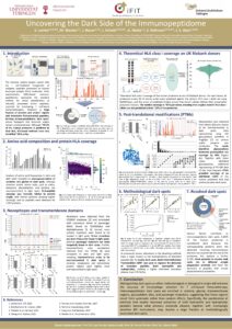
The immune system targets cancer cells via T cell-mediated recognition of antigenic peptides presented on human leucocyte antigen (HLA) molecules. Mass spectrometry (MS)-based immunopeptidomics represents the standard method for direct identification of naturally presented tumor peptides, essential for developing T cell-based immunotherapies. However, a large fraction of proteins and protein regions lack detectable HLA-presented peptides, forming immunopeptidomic ‘dark spots' whose biological and technical origins remain uncharacterized. Here, we used the PCI-DB, a uniquely large database comprising >10 million HLA-presented peptides, for comprehensive mapping of the immunopeptidome dark spot landscape. While HLA binding predictions suggest a median proteome coverage of 87% per person across UK Biobank donors, experimental immunopeptidome data have captured only 30% of the human proteome. HLA peptide coverage scales with gene expression, with the 11% of proteins lacking any detectable peptides showing the lowest expression. Analysis of cancer-related variants from the COSMIC database revealed that driver mutations are more frequent in light spots, whereas passenger mutations occur more frequently in dark spots. To investigate structural and post-translational contributors to dark spot formation, transmembrane annotations (TOPdb) and glycosylation sites (GlyGen) were compared to dark and light spots, revealing both to be overrepresented in dark areas. Reprocessing PCI-DB data to include glycopeptides increased glycosylation site coverage by 58%. Broader post-translational modification searches resolved ~1.4% of dark spots, with adapted MS acquisition methods expected to increase this further. Overall, characterizing dark spots as either technical or biological in origin will enhance epitope selection accuracy and expand the repertoire for T-cell-based immunotherapy.
Co-authors: Marcel Wacker, Jens Bauer, Jonas Scheid, Annika Nelde, Sven Nahnsen, Juliane S. Walz
Contact Attendee
Warning: Attempt to read property "user_email" on string in /home/1276969.cloudwaysapps.com/ydbgzhdjeq/public_html/wp-content/plugins/my-conference-now/functions.php on line 2826